Hip · Dr. Tania Ferguson

Abductor tears: gluteus medius and minimus tendon injuries

Pain on the outside of the hip is often called trochanteric bursitis, but the usual cause is injury or tearing of the gluteus medius and minimus tendons. Dr. Ferguson calls these muscles the rotator cuff of the hip, and treats them from the first round of therapy to complex revision repairs.
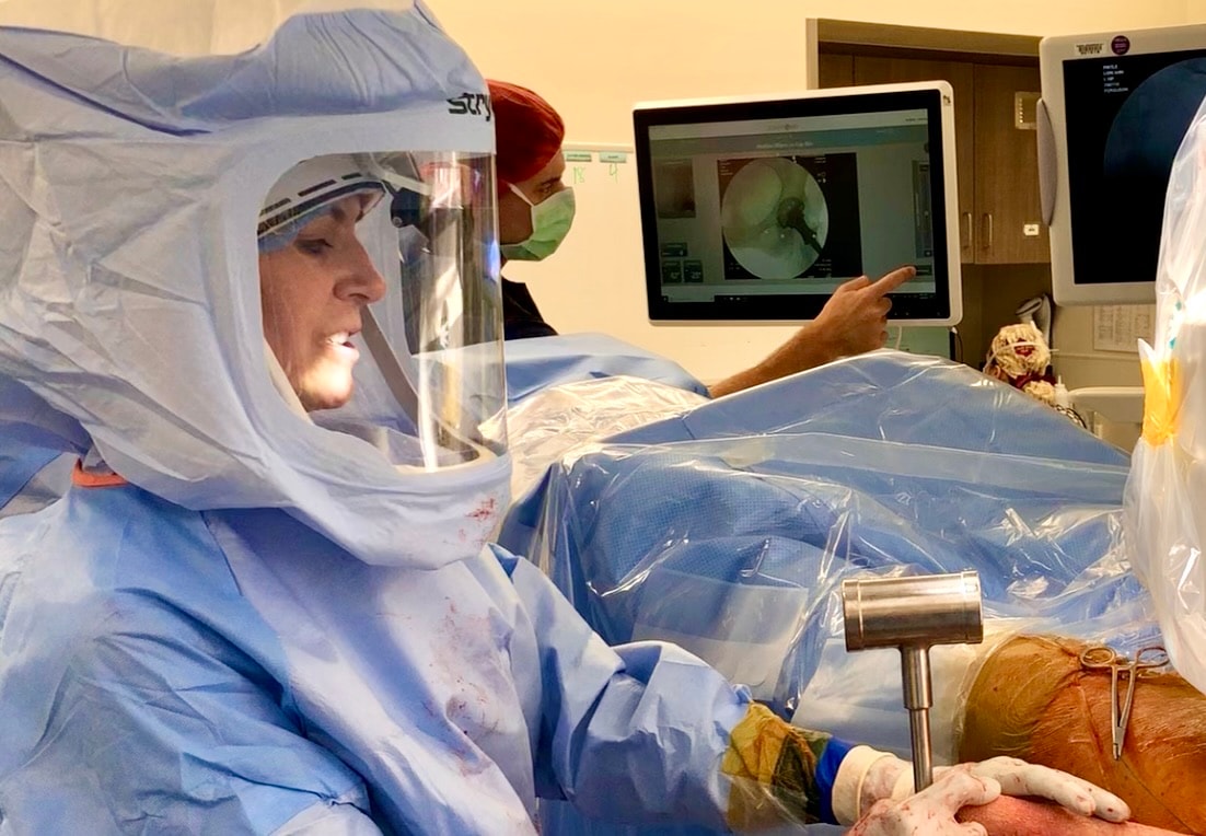
Dr. Tania Ferguson in a surgical helmet during hip surgery, as a colleague points to the live X-ray on the monitor

The evidence

The repair, in numbers

of Dr. Ferguson's abductor patients have had prior surgery that failed
50%More than
for the tendon to bond back to the bone as it heals
3–4Months
in the hospital for most patients after surgery
1Night

Symptoms

How an abductor problem feels

Patients with abductor problems feel pain on the outside of the hip, over the greater trochanter.

Pain on the side of the hip

The pain may radiate down the side of the hip to the knee.

Trouble with steps

Stepping up onto a step or a stool with the painful side is hard, as is walking up an incline. Many patients climb stairs leading with the other leg.

Pain lying on that side

Lying on the affected side hurts, often interrupting sleep.

Weakness and a limp

As you walk, the pelvis sways to the painful side (a Trendelenburg limp).

Often called bursitis

Bursitis is almost always the result of a tendon injury, not the primary problem.

What an abductor tear is

Pain felt in the side of the hip, or laterally, is often misdiagnosed as trochanteric bursitis. The common source of these problems involves injury, tendinitis, or tearing of the tendons of the hip's abductors (gluteus medius and minimus), which insert on the greater trochanter of the hip.

Abductor tendon problems range from irritation and inflammation of the tendons (tendinitis) to chronic breakdown of the tendon collagen (tendinosis), to partial and then complete tearing of the tendons from the bone. Tears can occur from an acute injury, but most often occur simply with overuse and degenerative changes with age. Dr. Ferguson commonly refers to the hip abductors as the rotator cuff of the hip. The injury patterns and treatments are quite similar.

Inflammation of the tendon commonly causes fluid to accumulate in the trochanteric bursa.
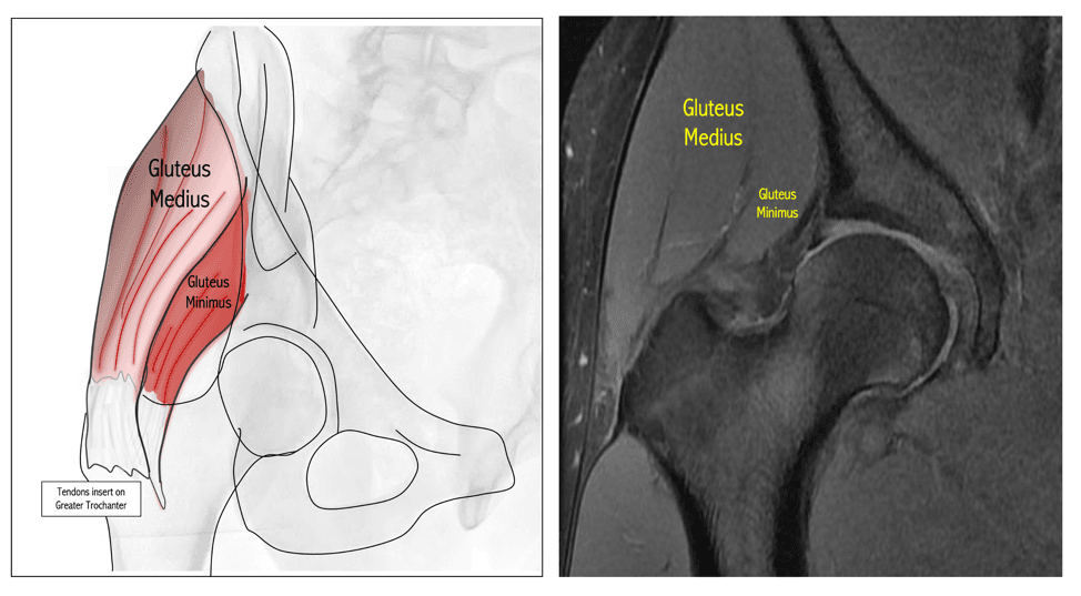
Left, a drawing of the hip showing the gluteus medius and gluteus minimus, with their tendons inserting on the greater trochanter. Right, an MRI of the hip with the same two muscles labeled
The gluteus medius and minimus tendons insert on the greater trochanter.

Non-surgical care

At the tendinitis stage, almost all patients respond to a coordinated conservative approach. Early abductor problems most commonly respond to activity modification, anti-inflammatory medications, and specific supervised physical therapy. Limited use of ultrasound-guided cortisone injections may help quell the pain of the tendon inflammation, and allow the physical therapy to work.

We do not endorse multiple, unguided steroid injections. They can lead to tendon attrition and poor blood supply, resulting in complete rupture and retraction of the tendon.

Physical therapy for abductor injuries includes a gentle, progressive strengthening program, and also muscle mobilization, dry needling, and collagen repair stimulating techniques that evoke a healing response in the diseased tendon. The majority of patients respond when the treatment is coordinated and the problem is caught early. An injection without a coordinated physical therapy program is not advisable, and commonly leads to more complicated tearing and repeated injections.

For patients who don't respond to the first round of conservative treatment, biologically active injections may be tried, including platelet rich plasma (PRP) and stem cells. These are thought to accelerate tendon healing.
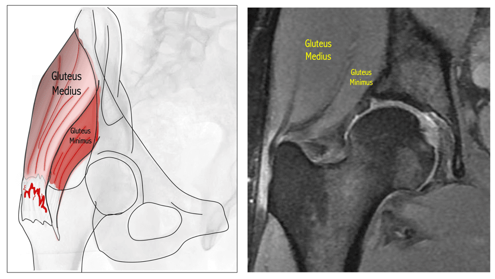
Left, a drawing of the gluteus medius and minimus with red marks of inflammation where the tendons insert on the greater trochanter. Right, an MRI of the hip with the two muscles labeled
Tendinitis at the insertion: inflammation, but no tearing, detachment or retraction. At this stage almost all patients respond to non-surgical care.

Surgical repair

Surgery to repair or reconstruct the tendon may be needed when non-surgical treatment has failed, or when the tendon has detached from the trochanter. Tears that are completely detached or retracted, have bone forming within the tendon, or have been operated on before most commonly need an open surgical approach. More than half of Dr. Ferguson's abductor patients have had one or more prior surgeries that failed.

Dr. Ferguson has developed a specific technique for abductor repair. As she puts it, "the damage found in surgery is virtually always far more extensive than identified with the MRI and ultrasound." The operation:
  1. 01Exposes the lateral trochanter and identifies the damage to the gluteus medius and minimus.
  2. 02Mobilizes the muscles, which are commonly retracted and scarred far from where they belong on the trochanter.
  3. 03Removes the scar tissue, fibrotic bursal tissue and bone fragments that fill the space where the tendon should attach.
  4. 04Frees the sciatic nerve from surrounding scar tissue, if it is encased, so it runs freely through the back of the hip.
  5. 05Clears the trochanter down to a healthy, bleeding bed of bone, so the tendon can heal back onto it.
  6. 06Reattaches the tendons to their normal footprint with several suture anchors, so the repair holds even if one anchor is lost during healing.
  7. 07Places a collagen-stimulating patch over the point of greatest damage to speed bone-to-tendon healing.
  8. 08Adds a silk augment where there is significant muscle atrophy, and in severe cases a stem cell preparation from bone marrow aspirate to help the muscle recover.
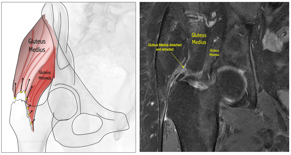
Left, a drawing of the gluteus medius and minimus with yellow points along the greater trochanter and arrows along the tendons. Right, an MRI labeled where the gluteus medius has detached and retracted
A gluteus medius tendon detached and retracted from the trochanter, drawn and on MRI.

Recovery, in four phases

The body takes three to four months to form the bond between tendon and bone. Recovery is planned in four phases around that.

  1. 01Weeks 0 to 8, protecting the repairWeight-bearing is limited to 30 lbs with the foot flat, with a walker or crutches for 10 weeks. No active abduction and no passive adduction. A hip abduction brace is worn out of bed, and a pillow between the legs in bed or in a chair. A high protein diet is recommended.
  2. 02Weeks 8 to 12The brace comes off and formal physical therapy starts. From week 10, weight-bearing increases under the therapist's direction until walking is limp-free without a walker, usually over two to four weeks. Pool walking and a stationary bike without resistance may start.
  3. 03Weeks 12 to 16Strengthening begins, and motion and weight-bearing restrictions are dialed back, with the goal of a normal, unassisted gait and pain-free hip motion.
  4. 04Week 16 onwardThe goal is a full return to normal activity. Any remaining weakness is treated with dry needling, neuromuscular electrical stimulation, laser therapy and guided strengthening.

Outcomes

Abductor tears are among the most painful conditions of the hip, and chronic tears often mean years of sleepless nights. Dr. Ferguson's practice sees these patients as the happiest surgical patients they treat. Many have almost no pain early after surgery.

The hardest part is often the period between four and eight weeks, when there is no pain but the restrictions still apply. Sticking with the rehab program is what makes the repair last. Recurrence can happen, particularly in hips operated on before and hips with severe muscle atrophy.

What to expect with surgery

  • Surgery commonly takes three hours or more, depending on the damage and scar tissue. Most patients spend one night in the hospital.
  • General anesthesia is used so the muscles relax fully for reattachment.
  • The incision is closed under the skin, with no stitches or staples to remove. A waterproof dressing stays on for seven days, then you change your own dressing every other day for two more weeks.
  • Aspirin daily for three weeks and compression stockings help prevent blood clots. When bone marrow aspirate is used (about one in ten patients), a different blood thinner and pain medication are used instead.
  • A GameReady cold compression device is recommended to reduce swelling. You'll get information about it before surgery.
  • A walker or crutches limit weight on the hip for 10 weeks, and a hip abduction brace is fitted at your pre-op appointment and worn out of bed for 8 weeks.
  • Formal physical therapy usually waits until week 8, saving your visits for the phase when hands-on therapy matters most.
  • Follow-up appointments and therapy protocols are laid out in the handouts on this page, and reviewed with you before surgery.

Your surgeon

Dr. Tania Ferguson

MD, MAS

Dr. Ferguson is a surgeon focused exclusively on the hip and pelvis. She trained under Joel Matta and Jeffrey Mast, two of the world's foremost experts in open hip surgery.

  • Recognized worldwide for hip preservation and acetabular reconstruction
  • Teaching the anterior approach to surgeons since 2005
  • Founding member, Anterior Hip Foundation
  • Trustee, AO Foundation
Dr. Tania Ferguson standing by a brick wall in her practice

Talk to Dr. Ferguson about your hip.

Request an appointment